Morphological changes of pseudoerosion
Morphological
changes of pseudoerosion- Patological anatomy
Supervisor
of studies - candidate of medical science Darjanova K.B., Yermenova
K.K.Kazakhstan Marat Ospanov State Medical University.K.Bekova
Aktobe
city, Republic of Kazakhstan
pseudoerosion of neck of uterus is
one of frequent a disease among diseases of neck of uterus [1].
It happens owing to independent recovery
congenital or the real erosion. Sometimes the pseudoerosion happens
recurrent, after the carried - out treatment. The long pseudoerosion of neck of
uterus leads to a hyperactivity of bazal cells, to complicitions of
proliferative activity, and it leads to
development of atypical cells, that leads to a dysplasia of necks of uterus
[2]. As a result of heavy form of a dysplasia the pseudoerosion develops in
precancer .
Objective:
. Identification
frequency of distribution of a pseudoerosion of neck of uterus
for the last 3
years (2010-2012
years) according to bureau
of the Aktobe regional
patological anatomy.
. Stydying of clinico
- morphological types of pseudoerosion of neck of uterus.
. Stydying of
a age features.
Method of research and materials:
Research was conducted with registry magazines of bureau of the Aktobe regional
patological anatomy for the last 3 years (2010-2012
years) and biopsy materials of it.
After fixation histological section with 10% formalin, is run out paraffin,
hematoxylin and eosine is painted standartly. The description of
micropreparations was carried - out by means of Leica
DM - 1000 microscope.and
analysis: The
pseudoerosion of neck of uterus among disease of neck of uterus - 2010-2012 years
- 1355, 2010 year -
572 (42,3%); 2011year -
401 (29,5%); 2012 year -
382 (28,2%) . Page 1 .
 1. Frequency of
incidence of a neck of uterus for 2010-2012 years
1. Frequency of
incidence of a neck of uterus for 2010-2012 years
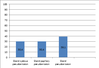
By the types of clinico
- morphological: gland
cystous pseudoerosion - 30,4%; gland
papillary pseudoerosion -
30,4%; gland
pseudoerosion -
39,1%. Page 2 .
 2 .
Frequency of incidence of neck of uterus on a clinico - morphological types for
2010 - 2012 years.
2 .
Frequency of incidence of neck of uterus on a clinico - morphological types for
2010 - 2012 years.
By the age features of pseudoerosion
of neck of uterus in reproductive
stage - 1170 (86,3%), premenapause -
135 (9,9%), menapause-
17 (1,2%), postmenapause -
33 (2,6%). Page 3.
 3. Frequency of
incidence of neck of uterus on a age features for 2010 - 2012 years.
3. Frequency of
incidence of neck of uterus on a age features for 2010 - 2012 years.
: glandulous cystous hyperplasia is
characterized by focal thickness of mucous membrane of cervical canal, glands
are covered with prismatic epithelial which produced mucus. Page 4.
 4. Glands are
covered with prismatic epithelial which produced mucus.
4. Glands are
covered with prismatic epithelial which produced mucus.
cystic widened glands with dense
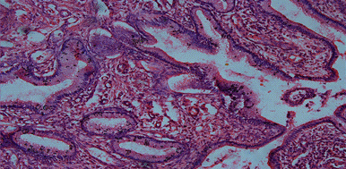
epithelium and partly swollen dense stroma may be observed.papillary
pseudoerosion of uterine neck the formation of papillary
outgrowths of cylindrical epithelium with eosinophili are observed. Page 5.
 5. Papillary
outgrowths.
5. Papillary
outgrowths.
glandulous hyperplasia a plenty of
differently - shaped structures covered by flatly or cubed epithelium with
eosinophil cytoplasm are seen. Page 6.
 6. Differently -
shaped structures.
6. Differently -
shaped structures.
 6. Flatly
celled metaplasia.
6. Flatly
celled metaplasia.
stroma is party swollen with a lot
of vessels and infiltration of leucocytes, lymphocytes and plasmacytes. Page 6.
 6. The stroma is
party swollen with a lot of vessels.
6. The stroma is
party swollen with a lot of vessels.
pseudoerosion neck uterus
The pseudoerosion of neck of uterus
meets in reproductive
ages,
between 16-46
age ( 1966 - 1996
years) more often and gland
pseudoerosion meets more often. The
reasaons of develop the disease: early sexual
life, damages at abortion or at the time of delivery, infections transmittable
sexual ways - clamidioses, trichomoniasis, mycoplasmosis, a virus of human
papilloma, and even to development from hormonal imbalances [3].
Uncomplicated ectopia of uterine neck is a variant of for female teenagers and
young women aged 20 - 25. For this age period ectopia of uterine neck
correlates to unstable hormonal status and as a rule doesn’t lead to
malignization and needn’t any treatment. But ectopia at the later age demands
careful examination and compulsory treatment. The risk of cancer is endangered
by combination of ectopia of uterine neck with inflammation processes of vagina
and uterine neck, leikoplakia of uterine neck, flat condyloma and pre - cancer
changes and dysplasia of uterine neck.
used
literature
1.
Аксель
Е.М. Статистика злокачественных новообразований женских половых органов. В кн.:
Клиническая онкогинекология . Руководство для врачей // Под ред. В.П.
Козаченко. - М.: Медицина, 2005. - С. 9-17.
.
Кондриков
Н.И. Патология матки. - Москва: Практическая медицина, 2008. - С. 28-72.
.
Хмельницкая
Н.М., Нейштадт Э.Л., Халимджанов З.К. Трудности и ошибки диагностики
атипической гиперплазии эндометрия // Арх. патол. - 2006. - №6. - С.39-42.